The interior organs of a human and its tissues may be seen clearly thanks to biomedical imaging methods. New perspectives on biological processes, such as modifications in receptor kinetics and molecular and cellular signalling, are made possible by today’s imaging techniques.
Biomedical imaging, which is the least intrusive, allows accurate tracking for the purposes of diagnosing, monitoring, and treating diseases. Sonography and ultrasound are included in this. The blog will discuss them while explaining sonography and ultrasound differences.
Learning about sonography and ultrasound
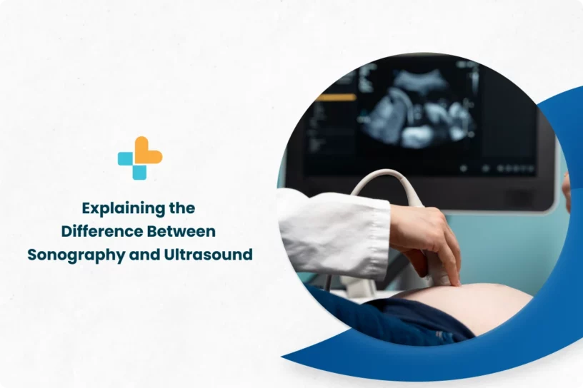
An ultrasound is one of the most popular imaging techniques, which uses sound waves to visualise the inner parts of the body (organs, tissues, or other structures). Unlike X-ray, which is another imaging method, ultrasound doesn’t employ the use of radiation, which makes it a safer choice.
Generally, ultrasound used for biomedical applications can be further classified into two types:
- Therapeutic ultrasound: This type of ultrasound is not used for imaging, but rather for treatment or therapeutic purposes. High-intensity sound waves are utilised to perform functions such as relocating or compressing tissue, heating tissue, breaking up blood clots, or giving medications to certain specific parts.
- Diagnostic ultrasound: Just as the name suggests, diagnostic ultrasound is used for diagnostic applications. It is a non-invasive technique and has quite high accuracy.
Sonography is a medical diagnostic process that creates powerful visual pictures of organs, tissues, or blood flow within the body using high-frequency sound waves (ultrasound).
How sonography and ultrasounds work
Most people have this question “how is sonography done?”. High-frequency sound beams are targeted onto the body which reflects back due to the organs and generates electrical signals. Now, these signals are processed by the computer to produce images.
Generally, a small probe-like structure, called a transducer, is used to send sound waves. The transducer is also responsible for receiving the echo of the waves sent inside. A few probes are also designed for entering the natural orifices (eg: rectum, vagina, oesophagus). This allows getting close to the organ or body part and getting a better view.
What is ultrasound used for?
Ultrasound may be used in a variety of settings. Maybe you’re expecting, and your obstetrician asks you to get an ultrasound to see how the baby is growing or to figure out when it will be due. Although, there is a difference between sonography and ultrasound in pregnancy, as explained in the next paragraphs.
Your doctor may have ordered an ultrasound to examine the blood flow because you’re experiencing issues with blood circulation in your heart or perhaps a limb. For many years, ultrasound has served as a popular imaging method in medicine.
What is sonography used for?
Are you wondering what a sonography test is ?. The sizes and forms of the internal body components can be seen on a sonogram obtained by sonography This diagnostic procedure is used to identify certain medical disorders by assessing the density, size, and form of tissue. It is the most effective method for seeing the abdomen without having to cut it open.
Doctors often opt for sonography before moving on to options such as CT scans and MRI, since both of them have complications and possible health risks.
Major differences between sonography and ultrasound
There aren’t many sonography and ultrasound differences. Sonography is the process, while ultrasound is the tool, to put it simply. Sonographers use ultrasound instruments to perform sonography. Ultrasound is the process of utilising sound waves to produce pictures from within the body, despite the terms being frequently used interchangeably. An ultrasound examination results in a picture known as a sonogram.
Limitations
The process of ultrasound has a few limitations, just like any other imaging technique. And they are:
- An ultrasound scan is a difficult option for obese people. The intra-abdominal fat prevents the sound waves from penetrating, thereby damaging the image quality.
- Because sound waves don’t propagate effectively through air or bone, ultrasonography is ineffective at imaging body areas like the brain and lungs that contain gas or are covered by bone.
- Unlike some other scans, an ultrasound image isn’t very detailed. For example, it cannot tell if a tumour is benign or malignant.
Conclusion
Ultrasound has major applications in the field of biomedical science. And most importantly, it’s completely safe and quite affordable. Now that the sonography and ultrasound differences consult the doctor are clear, it’ll be easy to make a decision.
At Ayu Health, one gets a professional diagnostic team and is experienced in conducting the imaging techniques as prescribed. Contact us at 636-610-0800 to avail the best services.
Our Hospital Locations
Gastroenterology Surgery Hospitals in Chandigarh | Gastroenterology Surgery Hospitals in Bangalore | Gastroenterology Surgery Hospitals in Jaipur | Gastroenterology Surgery Hospitals in NCR | Gastroenterology Surgery Hospitals in Hyderabad
Our Doctors
Gastroenterology Surgery Doctors in Chandigarh | Gastroenterology Surgery Doctors in Bangalore | Gastroenterology Surgery Doctors in Jaipur | Gastroenterology Surgery Doctors in NCR | Gastroenterology Surgery Doctors in Hyderabad
About the Author

Dr. S. Goel
Dr. S. Goel is a renowned Internal Medicine Specialist currently practicing at Ayu Health, Bangalore. He is a Specialist in Internal Medicine, Diabetes HTN, Paediatric Care, and Family Medicine.


