Introduction
Fetal echocardiography (echo test), or a fetal echocardiogram, is an ultrasound-like test that helps evaluate an unborn baby’s heart during the second trimester. This echo test for pregnant ladies is usually conducted between 18 to 24 weeks of pregnancy. The test provides a clearer view of the heart structure and function of the fetus to the doctor.
An echocardiogram is an exam that utilizes sound waves to visualize the fetus’s heart. The image obtained provides an insight into the formation and functioning of the baby’s heart. Fetal echocardiography also allows the doctor to observe the blood flow within the fetus’s heart, providing a detailed view to detect any irregularities in blood flow or heartbeat.
Why is this test important?
Fetal echocardiography is important as any abnormal result from this test can help in learning whether further testing is required. With a diagnosis, the doctor can assist the patient in managing the pregnancy and preparing for the delivery.
The test results can help the doctor in formulating a treatment plan that may be required post-delivery. Additionally, the doctor may also provide counseling to assist the parents with decision-making during the rest of the pregnancy.
Why Is a Fetal Echocardiogram Done?
A fetal echocardiogram is not a requirement for every pregnant woman, as routine prenatal ultrasound tests performed by the doctor can typically provide information on the proper formation of the fetal heart. A fetal echocardiogram is only necessary in cases where there is a concern for or higher risk of heart disease in the baby.
Some of the reasons that may require a fetal echocardiogram to be done include the following,
- A history of heart issues within the family
- A structural heart defect or a possible arrhythmia has been potentially detected through a routine prenatal ultrasound
- An abnormality in the baby’s chromosomes or genes has been discovered
- If multiple organ abnormalities, such as in the kidney, bones, or brain, are identified
- If the mother has taken some medications that may lead to Congenital Heart Defects, including antibiotics, anti-seizure, anti-depressant, and anti-inflammatory medications
- The mother has medical conditions like diabetes, connective tissue disease, or phenylketonuria
- In case the mother has rubella while being pregnant
- If the pregnant mother used alcohol or abused drugs
- In some twin pregnancies
Do I need to prepare for this procedure?
No preparation is required for the exam. In addition, unlike other prenatal ultrasounds, there’s no need to have a full bladder by drinking lots of water.
What happens during the exam?
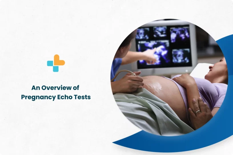
An echo test for pregnant ladies is similar to a typical pregnancy ultrasound. When the test is performed on the abdominal region, it is known as abdominal echocardiography. If the test is performed through the vagina, it is called transvaginal echocardiography.
- Abdominal echocardiography:
Abdominal echocardiography resembles an ultrasound, where the patient is asked to lie down and expose their belly. The ultrasound technician then applies a lubricating jelly on the skin, reducing friction and facilitating the movement of the ultrasound transducer, a device that sends and receives sound waves. The jelly also helps with sound wave transmission.
The transducer emits high-frequency sound waves through the body, echoing off the fetus’s heart. These echoes are recorded into a computer. The technician moves the transducer around the stomach to get images of the baby’s heart from different angles. Once done with the procedure, the jelly is removed from the stomach, and the patient can resume normal activities.
- Transvaginal echocardiography:
This echocardiography is often used during the early stages of pregnancy and can produce clearer images of the fetal heart. During a transvaginal echocardiography, the patient will be asked to undress from the waist down and then lie on an examination table. A technician will then insert a small probe into the vagina, which uses sound waves to create an image of the heart of the baby.
How Long Does a Fetal Echocardiogram Take?
A fetal echocardiogram can take between 30 minutes to 2 hours, during which the doctor will take images of the baby’s heart from different angles. In certain instances, it may take longer as the baby’s heart might be difficult to visualize due to the baby’s position.
Are there any risks associated with Fetal Echocardiogram?
Since a fetal echocardiogram does not use radiation, there are no known risks to the mother, or the baby are found to be associated with it.
What do the results mean?
Once the fetal echocardiogram is done, the doctor will explain the results to the patient in the next follow-up. If the results come out normal, it indicates that there were no cardiac abnormalities found.
If any problem, like an arrhythmia or heart defect, is found in the result, the doctor will prescribe further tests like a fetal MRI scan. The doctor may also prescribe a fetal echocardiogram again.
It is important to note that a fetal echocardiogram cannot be used for diagnosing every condition. For example, a hole in the baby’s heart cannot be detected with a fetal echocardiogram or even with highly advanced techniques. The doctor will inform the patient beforehand about the conditions that can be diagnosed with fetal echocardiogram and those that cannot be diagnosed.
Conclusion
There are various tests that are conducted during pregnancy to monitor the growth and overall well-being of the baby. One of the tests that are conducted between 18- 24 weeks during pregnancy is the fetal echocardiogram. This test is otherwise also called Fetal echocardiography. A fetal echocardiogram is an ultrasound-like test that helps evaluate the heart of an unborn baby during the second trimester.
At Ayu Health, we provide the best treatment, care, and other services needed during pregnancy. Our team of experts ensures that both mother and the baby get the best care throughout the pregnancy and post-delivery.
FAQs
1. what is the fetal echocardiogram (fetal echo test) price?
The price of a fetal echocardiogram may vary between INR 1200- INR 3500, depending upon the city, hospital, or laboratory.
2. Is fetal echocardiogram painful?
No, a fetal echocardiogram is not painful. However, you may experience pressure on the stomach or abdomen while the doctor moves the wand around it to get images from different angles.
Our Hospital Locations
Gynaecology Surgery Hospitals in Chandigarh | Gynaecology Surgery Hospitals in Bangalore | Gynaecology Surgery Hospitals in Jaipur | Gynaecology Surgery Hospitals in NCR | Gynaecology Surgery Hospitals in Hyderabad
Our Doctors
Gynaecology Surgery Doctors in Chandigarh | Gynaecology Surgery Doctors in Bangalore | Gynaecology Surgery Doctors in Jaipur | Gynaecology Surgery Doctors in NCR | Gynaecology Surgery Doctors in Hyderabad
About the Author

Dr. Nikitha Murthy B.S.
Dr. Nikitha Murthy B.S. is a renowned Gynaecologist currently practicing at Ayu Health, Bangalore.
He is s a Consultant with IVF Access at its Rajajinagar clinic. She has over 6 years of experience. Dr. Nikitha has a post-graduation (MS) in Gynaecology, DNB from the National Board of India, and a Fellowship in Reproductive Medicine. He also has vast experience in Post-Graduation (MS) in Gynaecology and DNB.


