ECG tests are life-saving because they can investigate symptoms of various heart problems.
While most people are familiar with the term “ECG”, not many know that hospitals employ many types of ECG tests to attain vital information on your heart’s health.
This article will help you understand how ECGs work, how many types of ECGs exist, the different types of ECG waves, and how doctors use them to save your life.
Let’s explore.
What is an ECG and How Does it Work?
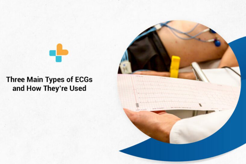
An electrocardiogram or ECG is a non-invasive, painless test that checks the rhythm and electrical activity of your heart. It works by detecting the electrical signals generated by the heart every time it beats using sensors attached to the skin.
These signals are recorded by a machine and then examined by a doctor to determine if they’re abnormal or not. Any irregularities detected may indicate a blockage, congenital anomaly, or weakness.
An ECG is usually recommended by cardiologists or other doctors who suspect the patient has a heart problem. A specially trained healthcare professional can perform the test in a hospital or clinic.
When is an ECG Used?
An ECG is usually used when symptoms of a heart problem, such as chest pain, palpitations, dizziness, and shortness of breath surface. It is frequently used in conjunction with other tests to help diagnose and monitor heart conditions.
An ECG can aid in detecting the following:
- Arrhythmias: This is a condition where the heart beats too slow, too quick or shows any irregular pattern.
- Coronary Heart Disease: This occurs when the blood supply to the heart is blocked or interrupted due to the buildup of fatty substances.
- Heart Attacks: When the blood supply to the heart is suddenly cut off, a heart attack occurs.
- Cardiomyopathy: Cardiomyopathy is a condition in which the heart walls thicken or enlarge.
What Are the Types of ECG?
Today, doctors use various types of ECGs to provide detailed information about a patient’s cardiovascular health. Given below are the three most common types of ECG machines.
Resting ECG
This is the most common type of electrocardiogram and the simplest to carry out. It’s performed when the person is lying still, hence the name resting ECG. It’s usually done as part of a routine checkup to detect heart conditions before any signs or symptoms appear.
The electrical activity of your heart is recorded from 12 electrodes on your chest, arms, and legs at the same time.
The patient is asked to lie down or sit up for 5–10 minutes for the duration of the test. The results will typically reflect your heart at rest.
Because of the number of electrodes used, it’s also called a resting 12-lead EKG.
Exercise ECG
Exercise ECG, also known as a treadmill test or stress test, is performed on the patient while they’re walking on a treadmill or pedaling a stationary bicycle. The intensity of the exercise is gradually increased while breathing rate and blood pressure are monitored.
This test is run to detect any coronary artery diseases or determine the safe exercise level for a patient after they’ve had a heart attack or heart surgery.
Patients are usually asked to fast and refrain from taking certain medications in order for this test to provide a clear and unbiased result. This makes sure the machine records the heart without any interference.
Holter Monitor
Holter monitor or ambulatory ECG is a type of electrocardiogram that’s used to continuously monitor the ECG tracking for a duration of 24 hours or longer.
The small plastic patches or electrodes are applied to specific areas. Then, the electrical activity is monitored, measured, and printed out for the doctor’s future reference.
The electrodes are linked to a small portable machine that’s attached to your waist, hence allowing you to monitor your heart at home for a day or more.
Types of ECG Waves
An electrocardiogram has three main components:
- The P-wave: It represents a small deflection wave that represents depolarizing atria.
- The QRS Complex: This represents ventricular depolarization.
- The T-Wave: It represents repolarizing ventricles.
Types of ECG Leads
There are three main types of ECG leads:
- Augmented Limb Leads (Unipolar)
- Limb Leads (Bipolar)
- Chest Leads (Unipolar)
The typical ECG has 12 leads. Six of them are referred to as “limb leads” because they’re attached to the person’s arms and legs. The remaining six leads are referred to as “chest leads” because they are located on the torso or chest.
The six limb leads called leads I, II, III, aVL, aVR, and aVF are the names of the six limb leads. These leads were initially named VR, VL and VF where V stands for Vector and R, L and F stands for Right, Left and Foot. A lowercase ‘a’ was later added which stands for ‘augmented’.
Leads V1, V2, V3, V4, V5, and V6 are the six precordial leads.
Understanding your ECG report may not be absolutely crucial but can help you understand the meaning behind clinical terms. It puts you in power to take care of your body and make the right choices for a healthy life. We hope this consolidated guide has helped you get a simplified yet comprehensive understanding of an ECG test and its types. At Ayu Health, we are committed to giving you the best healthcare. Contact us today to know more!
Our Hospital Locations
Cardiac Surgery Hospitals in Chandigarh | Cardiac Surgery Hospitals in Bangalore | Cardiac Surgery Hospitals in Jaipur | Cardiac Surgery Hospitals in NCR | Cardiac Surgery Hospitals in Hyderabad
Our Doctors
Cardiac Surgery Doctors in Chandigarh | Cardiac Surgery Doctors in Chandigarh | Cardiac Surgery Doctors in Bangalore | Cardiac Surgery Doctors in Jaipur | Cardiac Surgery Doctors in NCR | Cardiac Surgery Doctors in Hyderabad
About the Author

Dr. Magesh Balakrishnan
Dr. Magesh Balakrishnan is a renowned cardiologist currently practicing at Ayu Health, Bangalore.
He has 16 years of experience in this field. He has excellent skills in performing all cardiac diagnostic procedures/ tests. He has performed emergency and elective angiographies and angioplasties, device implantation (Pacemaker, AICD & CRT)


