While medical science has made enormous strides in the last decade, medical jargon can be quite intimidating to a layman.
This is especially seen in the case of heart-related jargon. In this regard, we’ll take a closer look at the two medical diagnostic tests — electrocardiogram (EKG/ECG) and echocardiogram (ECHO) — that are often recommended for heart patients by cardiologists.
Electrocardiograms (ECGs) and echocardiograms (ECHOs) are tests that can detect issues with heart muscles, valves, or the rhythm of the heartbeat.
This article will go through the difference between ECG and ECHO tests and how they can help you.
What Is an Electrocardiogram?
Also known as ECG or EKG, an electrocardiogram is a simple, non-invasive diagnostic test used to determine the rhythm and electrical imbalances in the heart. ECG machines are standard equipment in operating rooms and ambulances.
Why is an ECG Needed?
An ECG is needed to diagnose and monitor heart-related conditions. They’re used to look into symptoms that point to heart problems, like chest pain, heart palpitations, shortness of breath, and dizziness.
How Is an ECG Done?
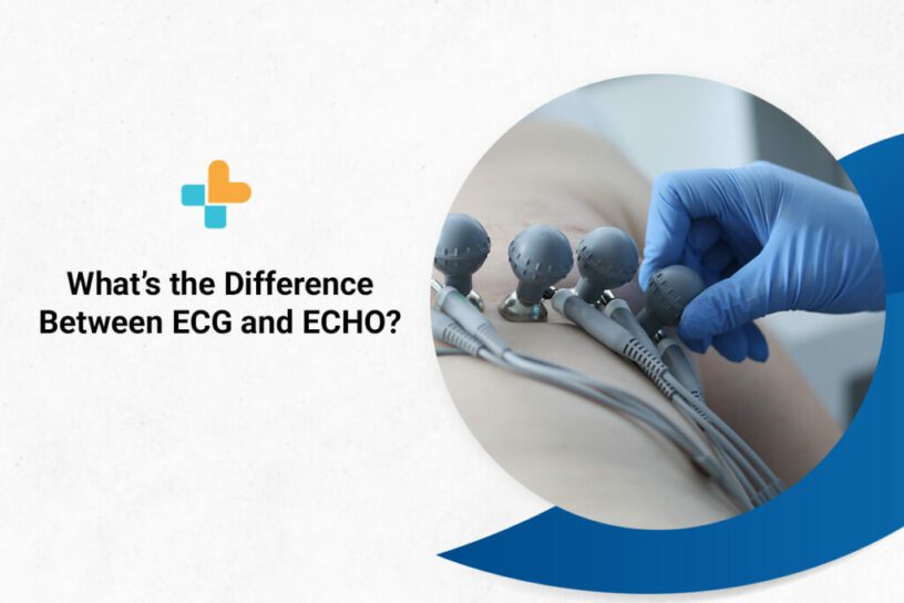
During an ECG, you’re made to lie flat on your back, and up to 12 electrodes (small adhesive sensors) are attached to your chest and limbs. These sensors are connected to a computer that keeps track of the electrical impulses that cause the heart to beat. The data is stored in the computer and displayed as waves on the monitor or paper.
You’re allowed to breathe during the exam, but you must remain completely still. Make sure you’re warm and comfortable before lying down. Moving, talking, or shivering may cause the test to fail.
An electrocardiogram (ECG) is a common and low-cost examination. They’re inexpensive and completely non-invasive (they don’t require needles or cuts). People with no signs of underlying heart conditions can take ECGs for simple check-ups.
You might even get one during a routine medical exam, especially if you have a close relative who has been diagnosed with heart disease. If you go to the emergency room with chest pain, palpitations, dizziness, difficulty breathing, or even simply overall weakness, your doctor may suggest an electrocardiogram.
Types of EKG/ECG
There are three main types of ECG.
- Resting ECG: This test is carried out when you’re lying down comfortably.
- Stress/Exercise ECG: This test is done when you’re on a treadmill or exercise bike.
- Ambulatory ECG (also called a Holter monitor): This test employs a small portable machine worn at your waist that monitors your heart for a day or more.
What Is an Echocardiogram?
Also known as an ultrasound scan, ECHO, or sonar of the heart, an echocardiogram is a non-invasive procedure that uses high-frequency sound waves to study the structure and functions of your heart and blood vessels.
These high-frequency sound waves, when emitted into the body, “echo” back, allowing you to observe the heart and all of its structures in real time.
Why Is an ECHO Needed?
An ECHO is needed to check how your heart chambers and valves pump blood throughout your heart. The results from an ECHO can help your doctor diagnose any potential heart conditions.
Being a non-invasive and affordable procedure, an ECHO is a go-to test among doctors for ruling out heart disease and other heart-related issues.
How Is an ECHO Done?
An ECHO is done by placing a transducer on your chest and aiming it at your heart. The transducer transmits and receives sound waves that bounce off the heart. A computer uses the bounced-off sound waves (echos) and turns them into a picture.
Types of ECHO
There are different types of echocardiograms available. Your doctor will request one based on the information they require. There are two commonly used ECHO tests.
- Transthoracic echocardiogram (TTE): Electrodes (small adhesive sensors) will be attached to your chest during the exam. These electrodes are connected to a machine that will measure your heartbeat. Your chest is then lubricated with a gel, and an ultrasonic probe is moved across it as you lie on your left side. A cable connects the probe to a nearby machine that will display and record the images it generates.
- Transoesophageal echocardiogram (TOE): Here, a smaller probe is passed down your throat into your food pipe (oesophagus) and sometimes into your stomach. The procedure is conducted using a local anaesthetic spray, and you’ll also be given sedatives to help you relax. You may need to fast for several hours before taking this test.
ECHO vs ECG
The following is a parallel comparison of the key features of an ECG vs echocardiogram to extend your grasp of the two jargon.
| EKG/ECG | ECHO | |
| Definition | Used to examine the electrical system of the heart. | Used to examine the mechanical system of the heart. |
| Result | The end result shows a wave-like diagram. | The end result shows a picture of the heart. |
| Duration and Procedure | It takes roughly 5 minutes to complete the test. The majority of that time is spent attaching the sensors; the recording itself only takes a few seconds to generate. | It takes roughly 20 minutes to complete. Five minutes will most likely be spent preparing for the procedure, and 15 minutes will be spent taking scans (pictures) of the heart. The test may take longer in some circumstances, depending on the exact information necessary. |
| Purpose | Detects irregular heart rhythm. Detects where the blood supply is blocked or interrupted within the heart. Monitors implanted pacemaker. Gives a baseline tracing of the heart’s function. | Detects problems with the valves or chambers of your heart. Detects birth defects that affect the normal working of the heart. Used to find the areas of damage or blockage in case of a heart attack. |
FAQs
Are ECG and ECHO the Same?
No, ECG and ECHO are not the same. An ECG (electrocardiogram) uses electrodes to look at abnormalities in your heart’s electrical impulses. An ECHO uses sound waves to determine irregularities in your heart’s structure.
Which Is Better: ECHO or ECG?
An ECHO is better than an ECG because they provide more accurate information on your heart valve functioning.
What Is an ECG and an ECHO?
An ECG/EKG (electrocardiogram) is a non-invasive test used to diagnose heart problems by determining the heart’s rhythm and electrical imbalances. An ECHO (echocardiogram) is a non-invasive procedure that uses high-frequency sound waves to study the structure and functioning of your heart and blood vessels.
Can an ECHO Detect Heart Blockage?
No, an ECHO cannot detect heart blockage. Your doctor might use a heart CT scan to find any calcium deposits or blockages in your heart arteries.
Can an ECG Detect Heart Blockage?
No, an ECG cannot detect a heart blockage. But it can detect the symptoms of a heart blockage. Your doctor might use a heart CT scan to find any calcium deposits or blockages in your heart arteries.
Why Is an ECHO Test Required?
An ECHO test is required to diagnose potential heart conditions. It’s used to determine how your heart’s chamber and valves are pumping blood.
Both these procedures are extremely important diagnostic methods for determining the cause of a wide range of heart issues. They’re both simple, non-invasive, and inexpensive when compared to other diagnostic tools.
We trust knowing the difference between ECG and ECHO tests will help you make the right decision for your next health check-up.
If your general healthcare practitioner recommends either of these tests, reach out toAyu Health to get the best care and services. With the top healthcare personnel and equipment, you can request our services in Bangalore, Chandigarh, Jaipur, and Delhi (NCR). Call us at +91 6366-100-800 for a consultation or book an appointment online!
Also known as ECG or EKG, an electrocardiogram is a simple, non-invasive diagnostic test used to determine the rhythm and electrical imbalances in the heart. ECG machines are standard equipment in operating rooms and ambulances.
An ECG is needed to diagnose and monitor heart-related conditions. They’re used to look into symptoms that point to heart problems, like chest pain, heart palpitations, shortness of breath, and dizziness.
Also known as an ultrasound scan, ECHO, or sonar of the heart, an echocardiogram is a non-invasive procedure that uses high-frequency sound waves to study the structure and functions of your heart and blood vessels.
These high-frequency sound waves, when emitted into the body, “echo” back, allowing you to observe the heart and all of its structures in real time.
An ECHO is needed to check how your heart chambers and valves pump blood throughout your heart. The results from an ECHO can help your doctor diagnose any potential heart conditions.
Being a non-invasive and affordable procedure, an ECHO is a go-to test among doctors for ruling out heart disease and other heart-related issues.
An ECHO is done by placing a transducer on your chest and aiming it at your heart. The transducer transmits and receives sound waves that bounce off the heart. A computer uses the bounced-off sound waves (echos) and turns them into a picture.
No, ECG and ECHO are not the same. An ECG (electrocardiogram) uses electrodes to look at abnormalities in your heart’s electrical impulses. An ECHO uses sound waves to determine irregularities in your heart’s structure.
An ECHO is better than an ECG because they provide more accurate information on your heart valve functioning.
An ECG/EKG (electrocardiogram) is a non-invasive test used to diagnose heart problems by determining the heart’s rhythm and electrical imbalances. An ECHO (echocardiogram) is a non-invasive procedure that uses high-frequency sound waves to study the structure and functioning of your heart and blood vessels.
No, an ECHO cannot detect heart blockage. Your doctor might use a heart CT scan to find any calcium deposits or blockages in your heart arteries.
No, an ECG cannot detect a heart blockage. But it can detect the symptoms of a heart blockage. Your doctor might use a heart CT scan to find any calcium deposits or blockages in your heart arteries.
An ECHO test is required to diagnose potential heart conditions. It’s used to determine how your heart’s chamber and valves are pumping blood.
Our Hospital Locations
Cardiac Surgery Hospitals in Chandigarh | Cardiac Surgery Hospitals in Bangalore | Cardiac Surgery Hospitals in Jaipur | Cardiac Surgery Hospitals in NCR | Cardiac Surgery Hospitals in Hyderabad
Our Doctors
Cardiac Surgery Doctors in Chandigarh | Cardiac Surgery Doctors in Chandigarh | Cardiac Surgery Doctors in Bangalore | Cardiac Surgery Doctors in Jaipur | Cardiac Surgery Doctors in NCR | Cardiac Surgery Doctors in Hyderabad
About the Author

Dr. Magesh Balakrishnan
Dr. Magesh Balakrishnan is a renowned cardiologist currently practicing at Ayu Health, Bangalore.
He has 16 years of experience in this field. He has excellent skills in performing all cardiac diagnostic procedures/ tests. He has performed emergency and elective angiographies and angioplasties, device implantation (Pacemaker, AICD & CRT)


